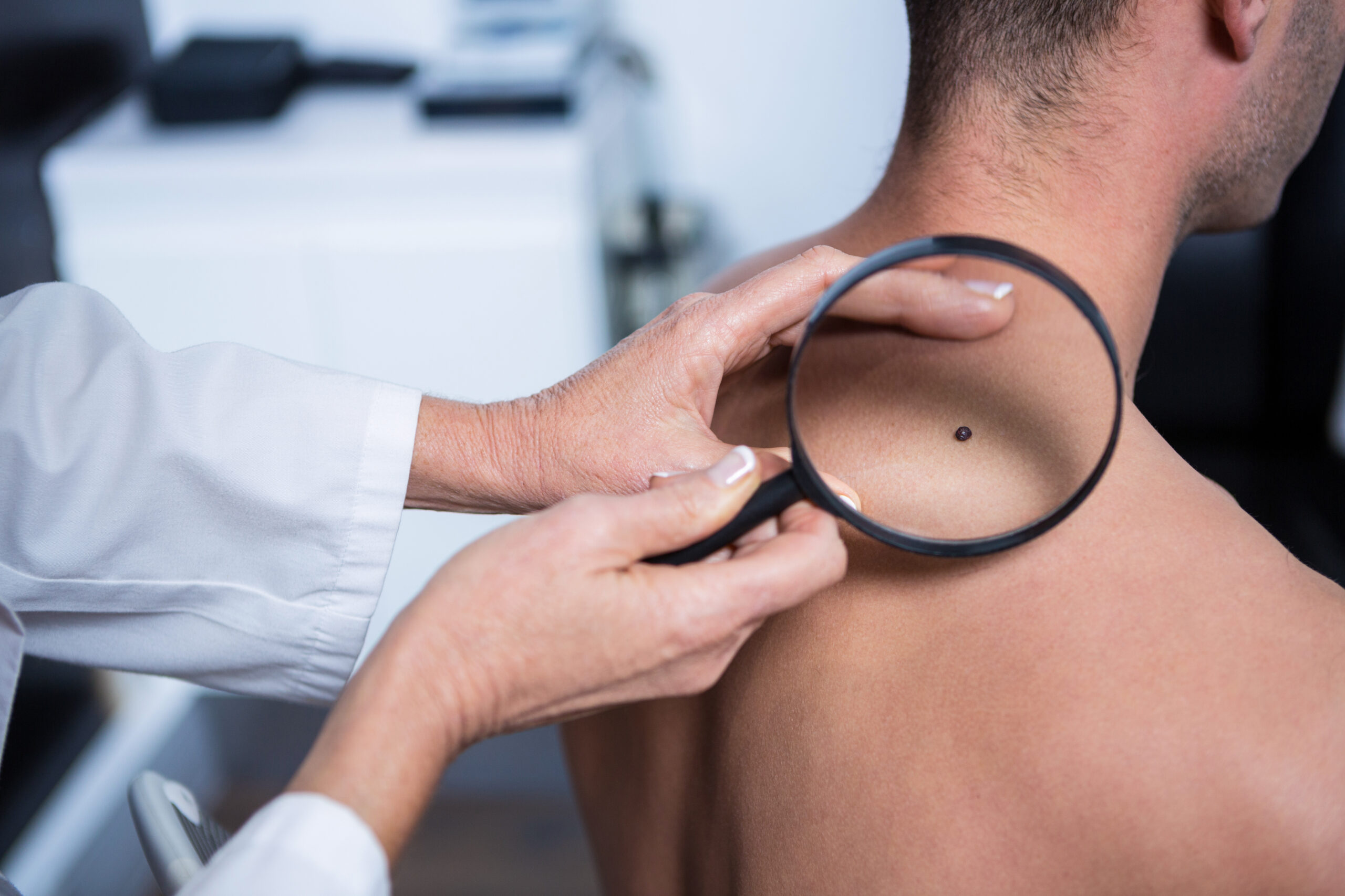
90% of the world’s population has moles. For some people, these are single small specks, for others – a whole stellar scattering. Nevi (as doctors call moles) can be of different sizes, shapes, colors and are found on absolutely any part of the body. And although they are classified as safe neoplasms, under the influence of unfavorable factors, they can degenerate into a malignant tumor or so called cancer.
Together with an expert, we will figure out what moles are, why they appear, and whether everyone should be afraid of cancer. who has them.
WHAT ARE MOLES
The outer skin layer (epidermis) is home to huge populations of cells called melanocytes. They protect our body from the negative effects of ultraviolet rays by producing the pigment melanin. It serves as a kind of sunscreen – it absorbs light waves and covers the skin with a tan. At the same time, dark-skinned people have much more melanin than light-skinned people (therefore, the latter burn out more easily). Melanocytes are very mobile, and if, when moving, they accumulate in one place, then a mole appears.
Nevi can be different in size, shape and color, both in the form of a small spot on the skin, and a giant lesion in size. The shade depends on the depth of the accumulation of melanocytes. Moles that are close to the surface have light brown, blue ones that lie very deeply, and dark brown ones in the interlayer between them.
Some nevi appear at birth, but for most, the reason is excessive exposure to ultraviolet radiation, hormonal changes in the body (puberty in adolescence and pregnancy) or various inflammations on the skin. Heredity also affects the appearance of moles: more moles will be in people whose mom, dad, grandparents had many nevi. There is a theory that some moles are transmitted at the genetic level, but there is no hard evidence for this.
Normally, a person has from 10 to 50 nevi, although in some of them their number can reach 400. At the same time, the number of moles is unstable, increasing by the age of 30–40, after which there is a tendency to decrease. At the age of 70–80 years, nevi may disappear altogether.
Moles should be distinguished from age spots, which include freckles, chloasma, and lentigo. In medicine, they are considered diseases (hyperpigmentation), since they occur when there is a malfunction in the production of melanin – melanocytes produce more of it than necessary. The causes of this disorder can be genetic or acquired (for example, due to health problems). So, a kind of age spots, inherited, are freckles. They are constantly present on the face, arms or shoulders, but they are activated only in spring and summer under the influence of increased sunlight. But chloasma and lentigo (brown spots of a bizarre shape of different shades) are acquired pigmentation on the skin. They usually occur due to abnormalities in the work of internal organs (inflammation, infections, hormonal disruptions), taking certain medications, or, more often, tanning abuse.
HOW ULTRAVIOLET LIGHT AFFECTS MOLES
Most skin cancers today are triggered by ultraviolet light. Every year the ozone layer is depleted, thereby increasing the amount of solar radiation to which people are exposed not only in hot countries, but also in northern latitudes. A burn can be obtained both in Sochi, Athens or Cairo, and in Stockholm, Reykjavik, Murmansk or Vorkuta. Constantly irradiated with ultraviolet light, melanocytes, one might say, fight against external influences – they produce melanin to protect the body. But their possibilities are not endless. At some point, they can fail. So, age spots are a kind of protest action on the part of the skin, which screams that all the limits of exposure to ultraviolet light have already been exhausted. The next level is skin cancer.
Why is ultraviolet radiation so harmful? The fact is that it stimulates mutations in D
NK. Some of them lead to the fact that cells begin to divide uncontrollably. Melanocytes can degenerate from benign to malignant and cause one of the most dangerous and rapid oncological diseases – melanoma. It is responsible for 80% of deaths caused by skin cancers. In this case, melanoma can be asymptomatic for a long time, hiding behind moles and age spots.
WHO IS MOST AT RISK
People with the first (red hair, fair skin and freckles) and the second (fair skin and hair) skin types are most at risk of developing this type of cancer. Nature has not endowed their melanocytes with the ability to produce melanin with a “high SPF”, as they were supposed to live in latitudes where there is little sun (in order to ensure the production of sufficient vitamin D, the skin must be more permeable to UV rays).
Also, people with the number of moles from 100 and above fall into the risk category. In most cases, nevi will not degenerate into anything bad. But the more there are, the higher the statistical probability. If your number of moles exceeds 50, it is important to undergo a full examination by a dermatologist, or even better, an onco dermatologist, once. The specialist will carefully check all moles, and if he confirms that none of the nevus causes concern, the next time you visit it is in 3-4 years. At the same time, no one cancels an independent examination. Those who have a family history of a malignant skin tumor should also be especially careful.
HOW TO CARRY OUT A HOME CHECKUP OF MOLES
Due to the fact that melanoma often hides behind nevi, it is important to regularly (ideally monthly) conduct a home examination of the skin for neoplasms. Especially if you are at risk. After all, timely detection of a malignant neoplasm promotes healing.
To check moles, you need to methodically study all parts of the body using two mirrors, not forgetting about the head (melanocytes are everywhere). Try to remember moles: watch the behavior of old ones and celebrate the appearance of new ones. In order not to miss an important sign, keep in mind the simple abbreviation AKORD:
A – asymmetry (a healthy mole is symmetrical and visually divided into two identical parts, a malignant one is not);
K – edges (in malignant – uneven);
O – color (non-uniform, for example, darker in the center and lighter at the edges);
Р – size (diameter is more than 4–6 mm);
D – dynamics (a malignant mole can change in size, shape, texture, wounds can appear on it).
If on examination it turns out that at least one mole has a sign of malignancy, be sure to go to the doctor.
image sources
- Dermatologist examining mole with magnifying glass in clinic: License Date: April 19th, 2023 Item License Code: 9ZTB2UCJXA


